


Lighting protein: Green Fluorescent Protein (GFP)
This page is for learning the structure and function of GFP by using the papaermodel of Green Fluorecent Protein (GFP).
1. What is GFP? - Intruduction of the molecule
Green fluorescent protein (GFP) was first isolated from Aequorea victoria, a kind of jellyfish living in the cold waters of the North Pacific, at the beginning of 1980's. The jellyfish contains a bioluminescent protein--aequorin--that emits blue light. GFP converts this to green light, which is what we actually see when the jellyfish lights up. In the 1990's, the method to express the GFP gene in other organisms such as worm, and the barrel shape structure of it was solved. 海洋生物におけるGFPの機能については今でも推測の域を出ていませんが、生物学の分野では様々な用途が見つかりました。一例は、バイオマーカーです。GFPなどの蛍光タンパク質を使うと、ある細胞や生物において特定の遺伝子がどこで発現しているのかをうまく追跡できます。
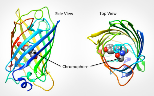
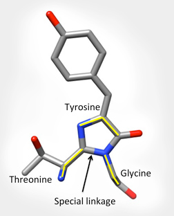
GFP分子の中で蛍光を担っている部分は発色団(chromophore)と呼ばれています。下図左では灰色、青、赤の球で示した、たるの中心にある部分がそれです。GFP分子内にある3つの特定アミノ酸残基(65番スレオニン-66番チロシン-67番グリシン)の主鎖原子が特別な結合を作り、環状型の構造(下図右)になります。このタンパク質が青色光を吸収し、緑色光を発する上でこの環状化は重要です。
より詳しくは「今月の分子 - 緑色蛍光タンパク質(GFP)」(英語原文・日本語訳)をご覧ください。
|
|
| 緑色蛍光タンパク質(GFP、PDB:1EMA) | GFPの発色団 |
2. GFPのペーパーモデルを作る
下記画像のリンク先はGFPペーパーモデルのPDFファイルです。これを印刷してモデルを作ってみてください。作る過程でタンパク質の一次構造、二次構造、三次構造が分かるでしょう。

- A4サイズPDF
- A4サイズトンボつきPDF(印刷業者への入稿用)
PDB 1ema: The information page of this structure (Yorodumi)
作り方を解説した動画もあります(英語)
関連情報
- Paper Model of Green Fluorescent Protein (GFP)…RCSB PDBの教育コンテンツサイト「PDB101」にある上記PDFの翻訳元(英語)
- Learning Resources: Green Fluorescent Protein (GFP)…このページの翻訳元。RCSB PDBの教育コンテンツサイト「PDB101」内(英語)
- 今月の分子 - 緑色蛍光タンパク質(GFP)(英語原文・日本語訳)
- 光るタンパク質 GFP - 万見プライム - PDBj
- 緑色蛍光タンパク質 - ウィキペディア